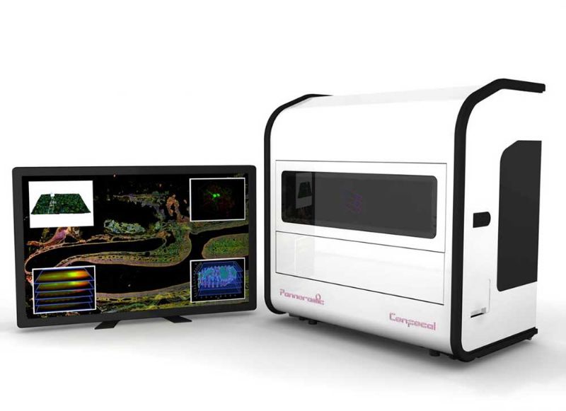
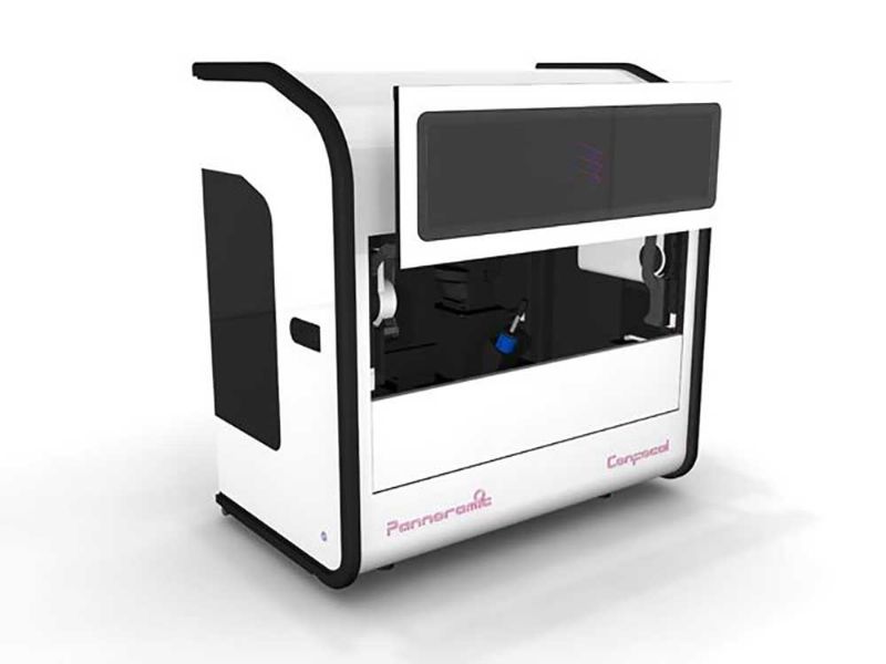
PANNORAMIC Confocal
PANNORAMIC Confocal offers confocal scanning for research pathology applications in unprecedented image quality and at unparalleled speed.
Research laboratories require maximum image quality (with low bleaching and phototoxicity) at a considerable scanning speed. While competitive technologies have not been able to deliver this, PANNORAMIC Confocal from 3DHISTECH offers fast and high-quality confocal scanning for molecular pathology applications by combining confocal imaging with award-winning whole-slide scanning technology.
Thanks to its innovative imaging technology, PANNORAMIC Confocal offers brightfield and fluorescence confocal scanning in unprecedented quality and speed for molecular pathology applications at low running costs, contributing to increased productivity for research laboratories.
Key Features
Innovative imaging technology
PANNORAMIC Confocal uses innovative structured illumination confocal imaging to overcome the limitations of spinning pinhole-disc techniques. This delivers the highest light efficiency with minimal bleaching and fastest scanning speed. Colocalized imaging is available with both fluorescent and brightfield illumination. Also, SW DDIC (Digital Differential Interference Contrast) is used to ensure low-contrast brightfield visualization.
Unique speed-up technologies
Technologies to increase scanning speed include darkfield and fluorescent preview, a Lumencor LED light engine for highest possible illumination, a scientific SCMOS camera combining high sensitivity and low noise to ensure short exposure times as well as an automated water immersion system for high NA objective. These solutions ensure a scanning speed up to 20 times faster than scanners using the spinning disc technology.
Anti-bleaching solutions
The structured illumination technology ensures minimum bleaching by collecting every usable light from the sample. Unnecessary sample illumination is guaranteed by hardware light triggering and reducible light intensity preserves the sensitive samples. The high-brightness confocal mode makes possible to detect weak signals without increasing the excitation light intensity.
Low operating costs
Compared to competitive technologies (laser scanning confocal and spinning disc), PANNORAMIC Confocal offers an unbeatable advantage in running costs: while competitive texhnologies use expensive lasers with low lifetime (1-2,000 hours), Confocal’s LED light engine has a lifetime of over 15,000 hours.
Advanced image handling
Last but not least, Confocal offers multiple image export options (ROI, grayscale/color, multichannel) as well as lossless export to third-party 3D applications.
The benefits of using PANNORAMIC Digital Slide Scanners
Specifications
| Slide capacity | 11+1 slides |
| Acceptable slide formats | 25.5 (+-0.5) mm x 75.5 (+-0,5) mm, 1(+-0.05) mm thickness |
| Default objectives | Zeiss Plan-Apochromat 20x/0.8 NA, Zeiss C-Apochromat (W) 40x/1.2 NA |
| Camera type | 5.5Mpx, 16 bit, low noise (1.3 e-) PCO edge cooled scientific CMOS camera |
| Image resolution (in focus plane) | 0,4 µm FWHM (with 40x 1.2NA objective) |
| Confocal sectioning | 1,43 µm FWHM (with 40x 1.2NA objective) |
| Fluorescent illumination | 6 channels Solid state light engine, 15000 hrs lifetime |
| Default fluorescent filter sets# filter cube positionsfilter type | Quad band: DAPI/FITC/TRITC/Cy5, 3 (BF+FL mode) or 4 (FL mode)single-/dual-/quad-band |
| Brightfield illumination | 3CCD equivalent separated R-G-B LED |
| Digital slide format | Proprietary digital slide format (MRXS) with lossless or JPG/JPGXR/JPG2000 encoding |
| Export opinions | single/multi channel |
| areafile format | annotation or whole slideTIFF/JPEG |
| Instrument dimensions W x D x H | 97 cm x 58 cm x 103 cm (39?x23?x41?) |
| Weight | 100 kg |
Scanner Software for Research
PANNORAMIC Scanners are powered by the PANNORAMIC Scanner Software for Research, which provides versatile, fast and high-quality slide scanning according to the imaging needs of cancer research laboratories, pharmaceutical companies or medical research institutes. Its key features include
- New Graphical User Interface (GUI)
- Scanning profiles for individual slides, multiple scanning locations and separate scanning of TMA slides for flexibility
- Several focus modes, new intensity stitching algorithm, two multilayer scanning options and further improved deconvolution for superior image quality
- Highly adjustable software and hardware settings
- Customizable user interface and scanning workflow
- Built-in slide conversion into the following file formats: DICOM, Tiled TIFF
Complementary Software Solutions
The 3DHISTECH software product portfolio is the most comprehensive one on the market. Whether it is tissue microarrays, IHC stainings, quantitative measurements, e-learning application and more, 3DHISTECH offers solutions for your research work.
Important Notice
Please note that the PANNORAMIC Confocal digital slide scanner is for research use only, and cannot be used for patient diagnosis or treatment selection.